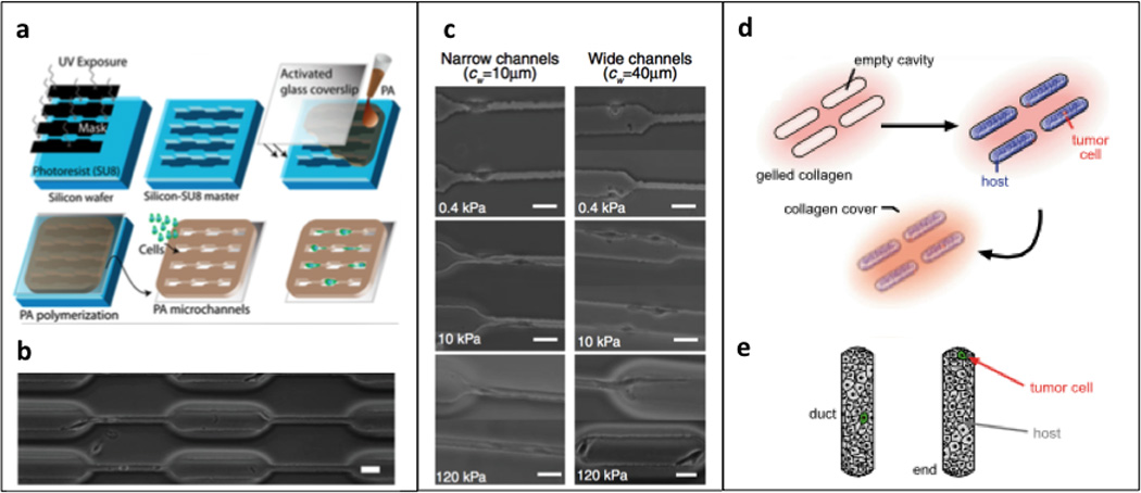
Figure 4. Microfabricated 3D substrates.

(a, b) Photolithography technique for generating polyacrylamide microchannels; reproduced with permission from [51]. (c) Phase contrast images of U373-MG human glioma cells migrating inside channels of varying stiffness and width, scale bar = 40 µm; reproduced with permission from [51]. (d) 3D microlithography approach to engineering mammary epithelial duct tissue; reproduced with permission from [53]. (e) schematic depicting “duct” versus “end” locations in epithelial host tissue; reproduced with permission from [53].
