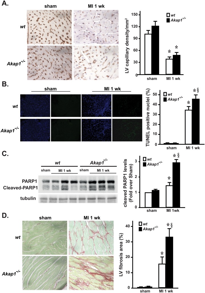
Fig 2. Akap1 genetic deletion enhances apoptosis and fibrosis after myocardial infarction.
(A) Left: Representative lectin staining of cardiac sections from wt or Akap1-/- hearts after the sham procedure or 7 days of permanent coronary artery ligation (MI 1wk). Capillaries appear brown. Right: Bar graphs of cumulative data of multiple independent experiments analyzing capillary density in the different groups (*p<0.05 vs. sham; n = 4 hearts/groups). (B) Left: Representative DAPI and TUNEL staining of cardiac sections from wt or Akap1-/- mice after the sham procedure or MI 1wk. Positive nuclei appear green. Right: Bar graphs of cumulative data of multiple independent experiments on TUNEL staining (*p<0.05 vs. sham; §p<0.05 vs. wt MI; n = 4 hearts/group). (C) Representative immunoblots (left) and densitometric analysis (right) of 4 independent experiments to evaluate cleaved PARP-1 protein levels in wt and Akap1-/- mice after the sham or MI 1wk procedures. Tubulin protein levels did not significantly change among the samples (*p<0.05 vs. sham; §p<0.05 vs. wt MI; n = 5 hearts/group). (D) Left: Representative images of Sirius red staining of cardiac sections from wt or Akap1-/- mice 7 days after the sham procedure or MI (60X magnification). Right: Bar graphs showing cumulative data of multiple independent experiments analyzing percent fibrosis in peri-infarct areas (*p<0.05 vs. sham; §p<0.05 vs. wt MI; n = 4 hearts/groups).

