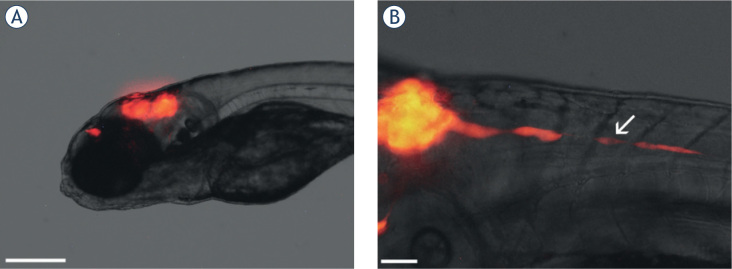
Figure 2.
In vivo imaging of glioblastoma cells in the brain of zebrafish embryos. (A) Embryo 3 days after the implantation of U87DsRed cells in the brain (visible as red fluorescence). Compact tumors have formed in the midbrain and for brain. (B) An embryo with implanted U87-DsRed cells 2 days after implantation, with a string of U87-DsRed cells rapidly invading from the tumor in the brain in the posterior direction (arrow). Scale bars: 300 μm (A); 50 μm (B).

