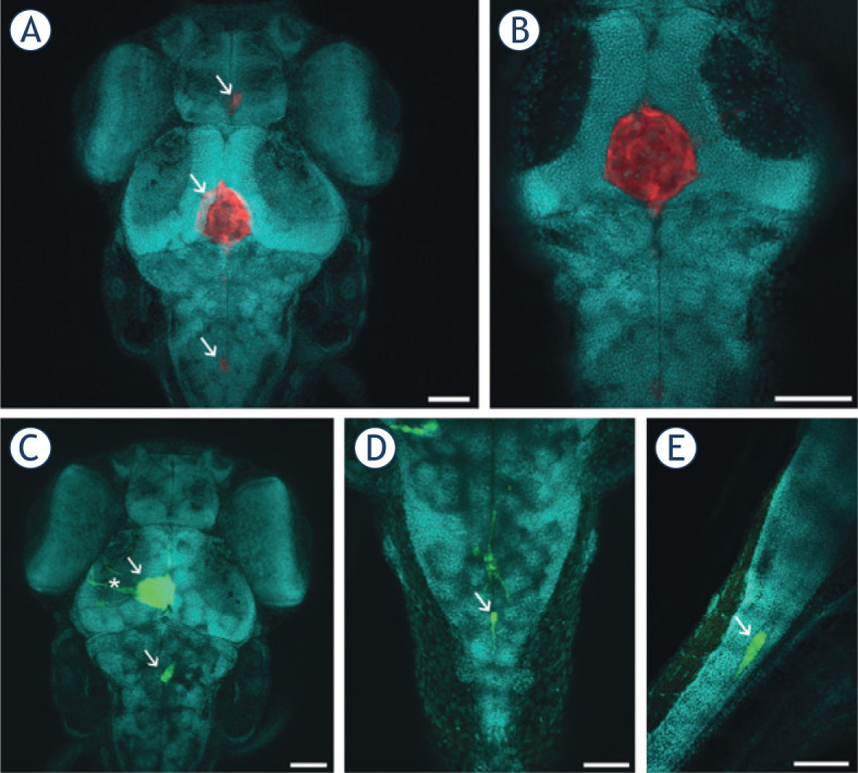
Figure 3.

Visualization of GBM cells in cleared zebrafish embryos, counterstained with methyl green (presented in blue). Embryos with U373-GFP and U87-DsRed cells implanted in the brain were cleared with ScaleU2, counterstained with methyl green, and imaged with confocal microscopy. (A) U87-DsRed cells (arrows) in the brain of a zebrafish embryo 3 days after implantation. (B) An optical section through the tumor in (A), demonstrating that the tumor is a compact mass of U87 cells in the midbrain ventricle. (C) U373-GFP cells (arrows) in the brain of a zebrafish embryo 3 days after implantation. Elongated U373-GFP cells are visible invading from the tumor (asterisk). (D) A U373-GFP cell invading along the central canal of the spinal cord (arrow), in dorsal view. (E) A U373-GFP cell invading along the central canal of the spinal cord (arrow), in lateral view. Scale bars: 70 μm (A, B, C); 50 μm (D, E).
