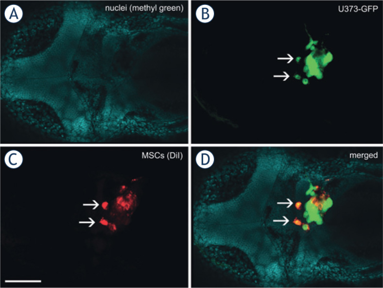
Figure 5.

Fusion between GBM cells and MSCs in the zebrafish brain. A mixture of U373-GFP cells and DiI-labeled MSCs was implanted in the brain of the embryos, which were fixed, cleared and imaged 3 days after implantation of the cells. Two cells (arrows) emit green GFP fluorescence as well as red DiI fluorescence, which strongly indicates that the U373-GFP cells and MSCs have fused after implantation.Nuclei of embryonic tissues labeled with methyl green. (B) Green fluorescent protein fluorescence of U373 cells. (C) Red fluorescence of DiI, used to label the MSCs. (D) Merged image of all of the fluorescent channels. Scale bar: 200 μm.
