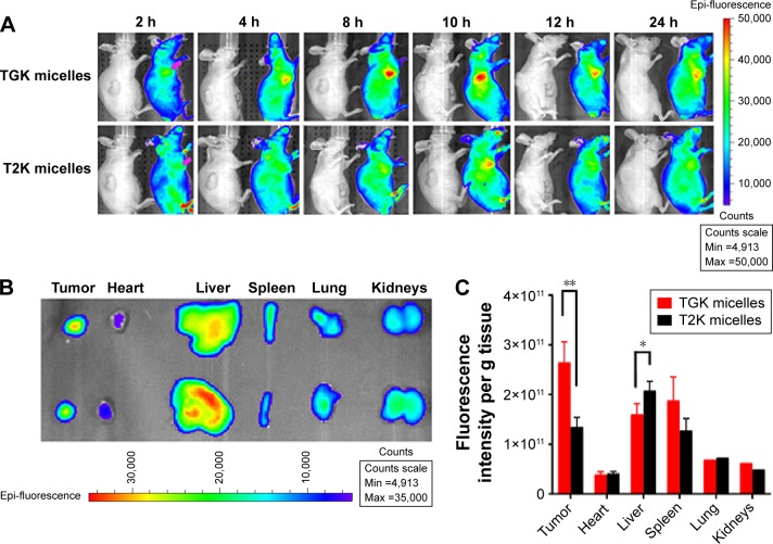
Figure 9.
In vivo imaging studies for tumor targeting ability of MMP-2/9 sensitive micelles.
Notes: (A) In vivo imaging of subcutaneous tumor-bearing nude mice after intravenous injection of DiR-labeled T2K micelles and TGK micelles at 2, 4, 8, 10, 12, and 24 hours postinjection, respectively. Tumor-bearing nude mice injected with saline (left of each group) were used as control. (B) Images of dissected organs of subcutaneous tumor-bearing nude mice executed at 24 hours after intravenous injection of DiR-labeled T2K micelles and TGK micelles. (C) Fluorescence intensity normalized with weights of DiR-labeled T2K micelles and TGK micelles in various organs. Values are expressed as mean ± SD (n=3); *P<0.05; **P<0.01.
Abbreviations: DiR, 1,1′-dioctadecyl-3,3,3′,3′-tetramethylindotricarbocyanine; DiR T2K micelles, DiR-labeled micelles composed of TPGS/T2K (n:n =40:60); DiR TGK micelles, DiR-labeled micelles composed of TPGS/TGK (n:n =40:60); SD, standard deviation; TPGS, d-α-tocopheryl polyethylene glycol 1000 succinate.

