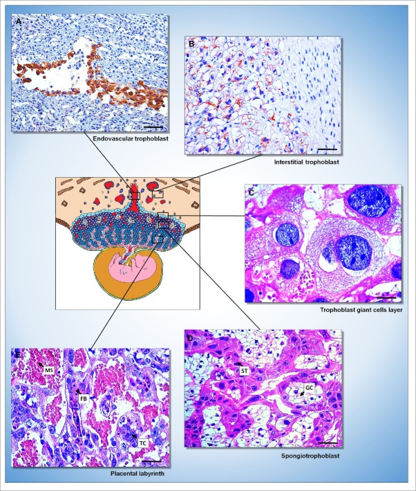
Figure 3.
Rat placenta. (A and B) Immunohistochemical evidence of the endovascular (A) and interstitial (B) trophoblast in the decidua stained with AE1/AE3 cytokeratin antibody. [Streptavidin-biotin-peroxidase method, Harris' hematoxylin counterstain, bar = 150 µm (A); 64 µm (B)]. (C-E) Histological evidence of the 3 layers of the placenta [trophoblast giant cells layer (C); spongiotrophoblast (D) formed by spongiotrophoblasts (ST) and glycogen cells (GC); placental labyrinth (E) formed by fetal blood vessels (FB), trophoblast cells (TC) and maternal sinusoids (MS)]. [Hematoxylin and eosin, bar = 64 µm].

