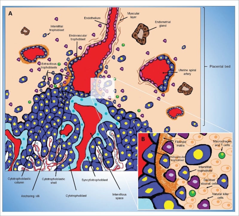
Figure 4.

Intrauterine trophoblast invasion in humans. (A) The placental villi are covered by villous trophoblast cells (an inner cytotrophoblast layer covered by syncytiotrophoblast). O core of villus contains fetal blood vessels, fibroblasts and fetal macrophages. Maternal blood in the intervillous space reaches the placenta through the uterine spiral arteries. Cytotrophoblasts leave the top of the anchoring villi and become extravillous trophoblasts. Initially, the extravillous trophoblasts form cell columns and then invade and migrate toward the decidua. The extravillous trophoblast residing outside of placental villi acquires the capacity to degrade extracellular matrix proteins to invade the decidua (interstitial trophoblast) and the wall of maternal uterine spiral arteries (endovascular trophoblast). Endovascular trophoblasts invade the lumen of the spiral arteries and incorporate into the arterial wall. The endothelium, the muscular layer and the elastic material of these arteries are destroyed and replaced with an extracellular matrix called the fibrinoid and by the EVT cells themselves. (B) Representation of the placental bed showing interstitial trophoblast cells between decidual stromal cells, uterine natural killer cells and others maternal leukocytes (macrophages and T cells).
