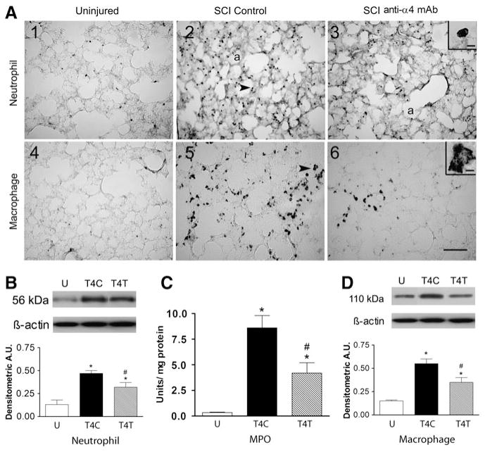
FIG. 1.
The anti-α4 treatment decreases neutrophils and macrophages in the lung at 24 h after spinal cord injury (SCI). (A) Photomicrographs of lung sections immunostained by an anti-neutrophil antibody (panels 1–3), and by an ED-1 antibody to detect macrophages (panels 4–6) from an uninjured rat, a T4 SCI control rat, and a T4 SCI rat treated with the anti-α4 monoclonal antibody (SCI anti-α4 mAb). The insets in A3 and A6 show high-power detail of stained cells (a, alveolus). The arrows in A2 and A5 point to a neutrophil and a macrophage, respectively (scale bar = 100 μm in A6 applies to A1–A6; scale bar = 10 μm in insets). (B) Neutrophil protein, identified by Western blotting in lung homogenates from uninjured and SCI rats (n = 4 for all groups) expressed in arbitrary units (A.U.; U, uninjured rats; T4C, T4 control SCI rats; T4T, T4 SCI rats treated with the anti-α4 mAb). A representative autoradiogram of a Western blot showing relative protein expression, compared to loading controls (β-actin), is shown above the bar graph. (C) Myeloperoxidase (MPO) activity in lung homogenates from uninjured rats (n = 6), T4 SCI control rats (n = 4), and T4 SCI rats treated with the anti-α4 mAb (n = 5). (D) Macrophage protein (ED-1) expression (Western blotting) in lung homogenates from uninjured and SCI rats (n = 4/group). In this and all figures values are means ± standard error (*significantly different from uninjured; #significantly different from SCI control; p ≤ 0.05 by Student Neuman-Keuls test for all comparisons).

