
FIG. 9.
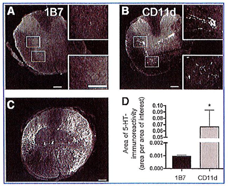
CD11d monoclonal antibody (mAb) treatment increases serotonin (5-HT) immunoreactivity caudal to the lesion. Immunohistochemistry was used to detect 5-HT in the spinal cord 42 days post-injury. Representative photomicrographs of sections stained for 5-HT immunoreactivity 1920 μm caudal to the lesion epicenter from 1B7- (A) or CD11d-treated (B) mice. (A) Almost no observable 5-HT staining was detected caudal to the lesion in 1B7-treated mice (5 mice/group). (B) Serotonin immunoreactivity was observed caudal to the lesion in the intermediolateral cell column and ventral horn in the injured spinal cords from animals treated with the CD11d mAb (6 mice/group). (C) Representative photomicrograph of a section from the lesion epicenter of a CD11d-treated mouse stained for 5-HT immunoreactivity. The appearance of the 5-HT immunoreactivity in the lesion epicenters of 1B7 mAb-treated controls was no different from the appearance of the 5-HT immunoreactivity seen in the CD11d mAb-treated mice (scale bars = 100 μm). (D) Measuring the area of 5-HT-immunoreactivity (area/area of interest) at 1920 μm caudal to the lesion epicenter in CD11d- and 1B7-treated spinal cord-injured mice revealed a significant increase in 5-HT immunoreactivity in the CD11d mAb-treated group.