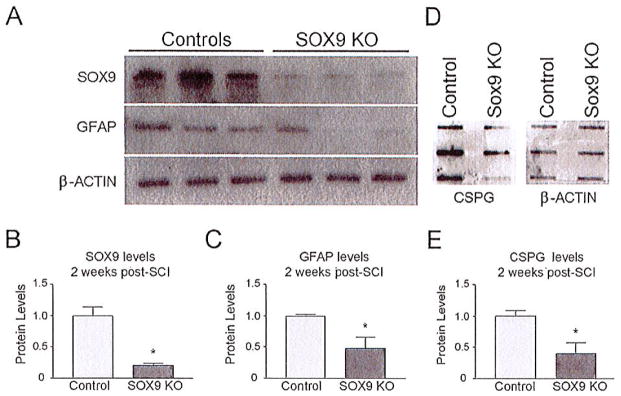
Fig. 3.
Sox9 conditional knock-out mice demonstrate reduced SOX9, GFAP, and CSPG protein 2 weeks post-SCI. (A) Western blot analysis and subsequent densitometry (B, C) demonstrate reduced SOX9 and GFAP levels in Sox9 conditional knock-out mice in comparison with control mice (normalized to β-actin levels) (P ≤ 0.05, Student’s t-test; n = 3). (D) Slot blot and subsequent densitometry (E) demonstrates reduced CSPG expression in Sox9 conditional knock-out mice compared with controls (normalized to β-actin levels) (P ≤ 0.05, Student’s t-test; n = 3).

