Fig. 5.
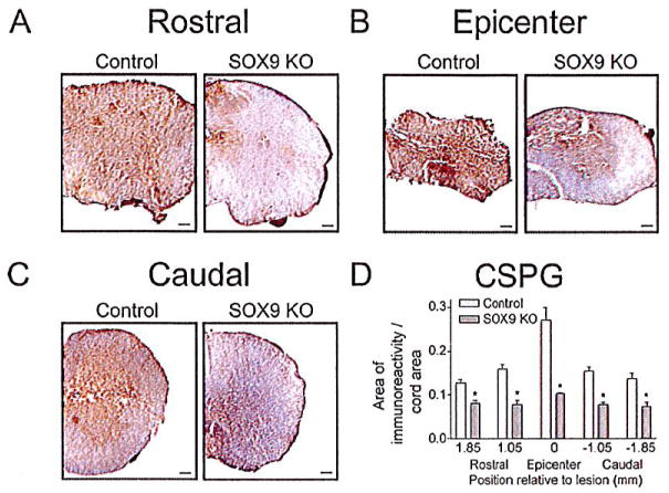
Sox9 conditional knock-out mice display reduced CSPG expression 14 weeks post-SCI. Representative photomicrographs of anti-CSPG DAB immunohistochemical staining approximately 1 mm rostral to the lesion epicenters (A) at the epicenters (B) and 1 mm caudal to the lesion epicenters (C) from Sox9 conditional knock-outs and controls as indicated. Bar = 100 μm. (D) Quantification of area of CSPG immunoreactivity in Sox9 conditional knock-out and control sections. The area of immunostaining per cross-sectional area of spinal cord was quantified using ImageProPlus software on sections spaced 160 μm apart. The area per area measurements were then grouped into bins centered on the positions indicated. The bin representing epicenter in each animal extended 0.65 mm rostral and caudal to the center of the lesion. The bins rostral and caudal to the epicenter were centered on the positions shown relative to the epicenter and included sections 0.4 mm rostral and caudal. *Statistically significantly different from controls (P < 0.05, two-way ANOVA, Newman-Keuls post hoc test (P < 0.05); n = 9 Sox9 KO, n = 11 controls).

