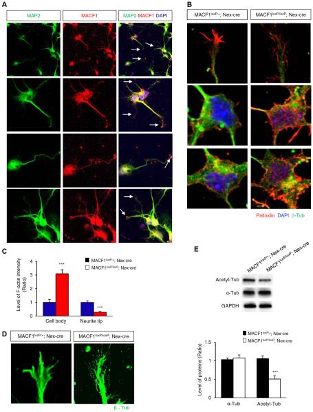
FIGURE 6. Actin and microtubule localization and arrangement in MACF1 mutant neurons.
(A) MACF1 expression in cortical neurons. Cortical neurons from E14.5 control (MACF1loxP/+; Nex-cre) and MACF1loxP/loxP; Nex-cre brains were cultured for 5 days, and MACF1 localization was examined using immunostaining. Arrows indicate an accumulation of MACF1 in dendritic tips (B) MACF1 deletion causes aberrant actin arrangement and localization. Polymerized actins were visualized by phalloidin staining. Top panels show the patterns of polymerized actins at neurite tips. Middel and bottom panels show polymerized actins accumulated in the cytosol. (C) The levels of polymerized actin (F-actin) intensity in the cell body and at the neurite tip were quantified using Image J (NIH) software. n = 21 cells from 3 independent cultures using 3 mice for each condition. Statistical significance was determined by two-tailed Student's t-test. ***p < 0.001. (D) MACF1-deleted neurons show abnormal microtubule arrangement at the neurite tip. (E) Immunoblotting was performed to measure the levels of α-tubulin or acetylated-tubulin using E14.5 control and MACF1loxP/loxP; Nex-cre brain lysates (top panel). The levels of each tubulin were quantified in the bottom panel. n = 3 blots using 3 different lysates from 3 mice for each condition. Statistical significance was determined by two-tailed Student's t-test. ***p < 0.001.

