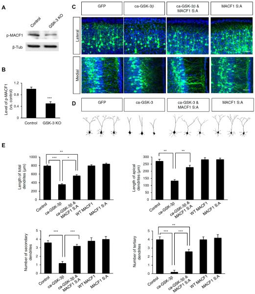
FIGURE 7. MACF1 mediates GSK-3 signaling in dendritic branching.
(A) GSK-3 deletion inhibited phosphorylation of MACF1in the developing brain. Phosphorylation of MACF1 was measured by Western blotting using brain lysates from control and GSK-3 knockout mice (GSK-3α−/−; GSK-3βflox/flox; Nestin-cre). Control: GSK-3α+/−; GSK-3αflox/+; Nestin-cre. (B) Quantification of (A). (C) Suppression of GSK-3 phosphorylation of MACF1 partially restores the inhibitory effect of GSK-3 in dendrite outgrowth. E14.5 mice were electroporated in utero with a GFP, ca-GSK-3β-GFP, WT MACF1, MACF1 S:A-GFP, or ca-GSK-3β-GFP and MACF1 S:A-GFP constructs. Brain sections were prepared at P14 to assess dendrite outgrowth and branching. The overexpression of ca-GSK-3β-GFP inhibited dendrite outgrowth. However, the defective dendrite outgrowth was partially rescued by co-overexpression of ca-GSK-3β -GFP with MACF1 S:A-GFP construct. Scale bar: 25 μm. (D) Representative single cell traces soma and dendrites shown in (C). (E) The lengths and numbers of total and primary/secondary apical dendrites were quantified. n = 75 cells from 5 mice for each condition. Statistical significance was determined by one-way ANOVA with Bonferonni correction test. *p < 0.05, **p < 0.01. ***p < 0.001.

