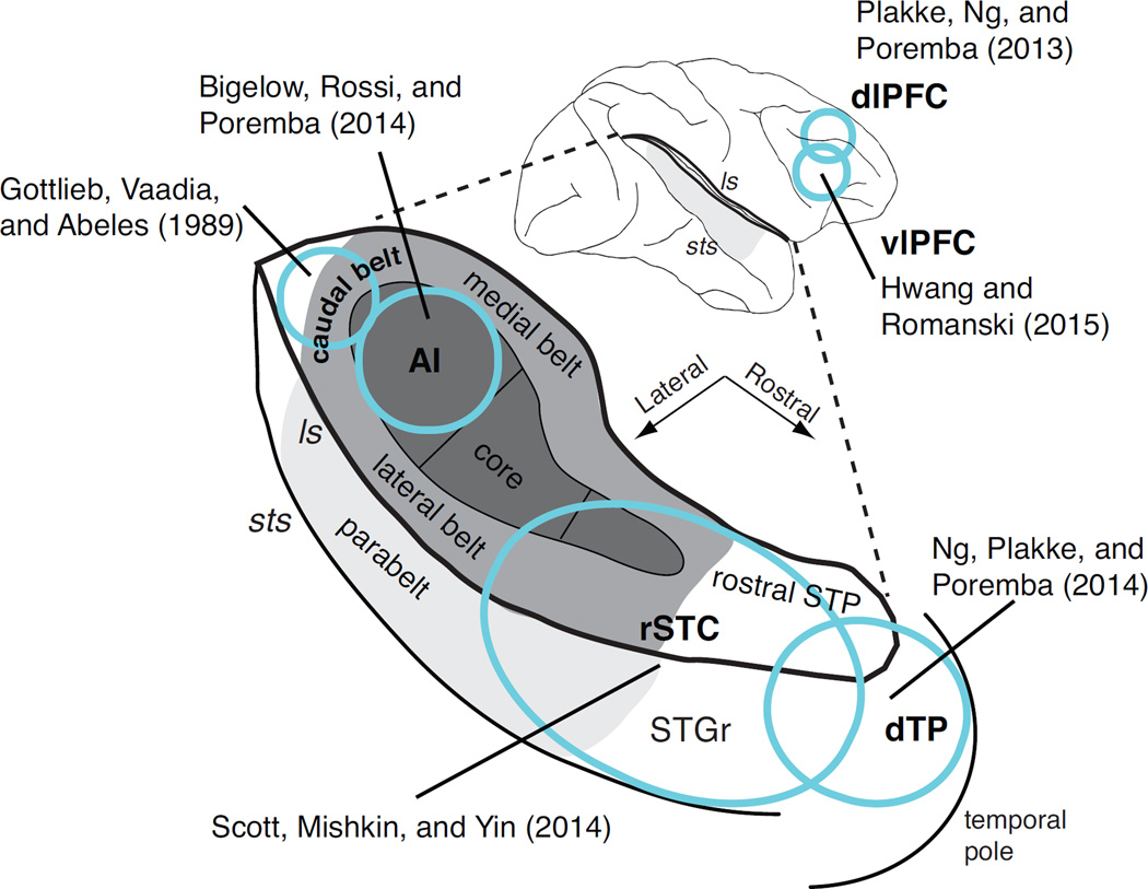
Figure 2.
Cortical regions recorded in neurophysiological studies of auditory STM in monkeys, collected on a schematic depiction of the macaque brain. Top, the bold black line highlights the lateral sulcus (ls), which has been opened up to reveal the auditory areas of the supratemporal plane (STP) illustrated below. The ascending hierarchical levels of core, belt, and parabelt are shaded from dark to light gray. Areas recorded in frequently cited studies are indicated in bold type and outlined in blue: the primary auditory cortex (AI, a subdivision of the core), caudal belt, rostral superior temporal cortex (rSTC), and dorsal temporal pole (dTP). Note that rSTC includes rostral core and belt, as well as the superior temporal gyrus (STGr), rostral STP, and some overlap with the dTP. The dorsolateral (dlPFC) and ventrolateral (vlPFC) prefrontal cortex is indicated on the lateral view at the top. A right-hemisphere view is used for convenience, but in actuality recordings were obtained from the right hemisphere in caudal belt (N=1 baboon; Gottlieb et al., 1989), one right and three left hemispheres in rSTC (N=3 rhesus monkeys; Scott et al., 2014), one left and two right hemispheres in vlPFC (N=2 rhesus, Hwang and Romanski, 2015; and the left hemisphere in AI, dTP, and PFC (N=2 rhesus; Bigelow et al., 2014; Ng et al., 2014; Plakke et al., 2013).

