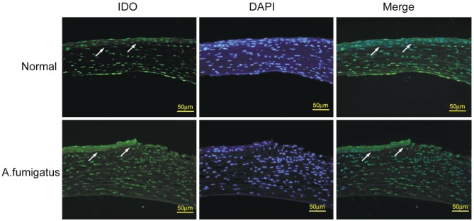
Figure 2. Immunofluorescence staining of IDO in the normal and A. fumigatus-infected corneas in C57BL/6 murine models.
Serial sections of normal and A. fumigatus-infected corneas at 3d were stained using IDO antibody and FITC-conjugated rabbit antimouse second antibody (for IDO, green), and counterstained with 4′,6-diamidino-2-phenylindole (DAPI) (blue). As indicated by the arrows in the normal cornea arrayed in the first row, few green fluorescence was observed in corneal epithelium. However, in the A. fumigatus-infected corneas (second row), strong green fluorescence in corneal epithelium was detected. Scale bar in immunofluorescence images: 50 µm.

