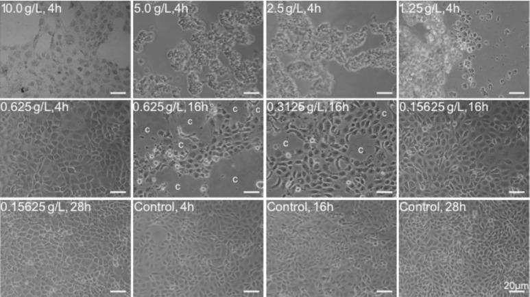
Figure 1. Morphological abnormality of Tetracaine-treated HCEP cells.
Cultured HCEP cells were treated with the indicated concentration and exposure time of Tetracaine, and their growth status and morphology were monitored by light microscopy. One representative photograph from three independent experiments was shown. c: CPE. Bar: 20 µm.

