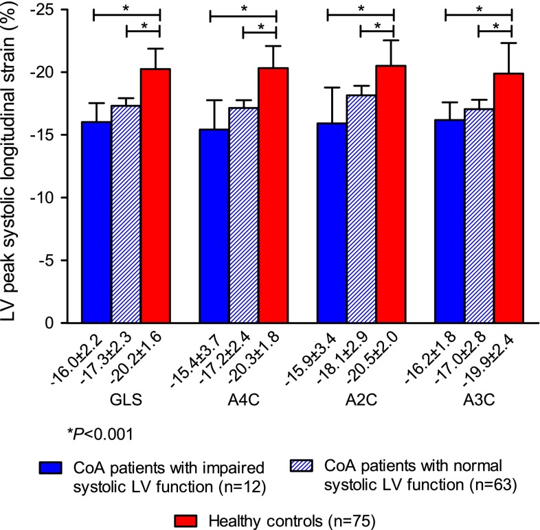
Fig. 3.
Left ventricular global longitudinal strain in CoA patients with visually graded impaired and normal left ventricular systolic function and in healthy controls. This figure presents the mean peak systolic LV GLS based on the measurements in the apical four-, two- and three-chamber view, and the mean peak systolic longitudinal strain at these three views separately. Strain values of the patients with visually graded normal left ventricular systolic function and of patients with impaired left ventricular systolic function are both compared with those of healthy controls. A2C apical two-chamber view, A3C apical three-chamber view, A4C apical four-chamber view, GLS global longitudinal strain, LV left ventricular

