. 2016 Apr 28;16(2):134–139. doi: 10.4110/in.2016.16.2.134

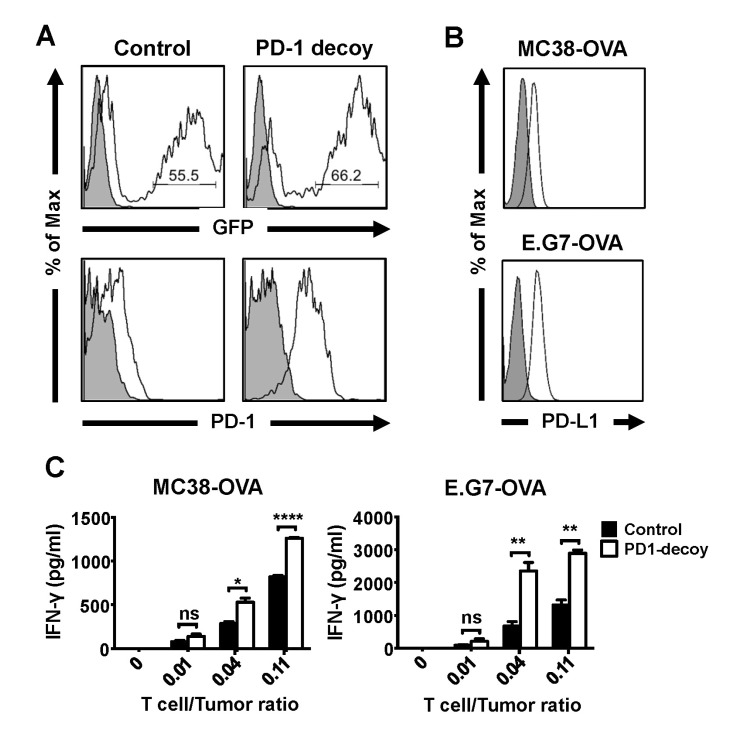
Figure 2. Enhanced anti-tumor reactivity of OT-I T cells carrying PD-1 decoy. OVA-specific CD8 T cells (OT-I cells) were transduced with retroviruses carrying control vector or PD-1 decoy as described in the methods and materials. (A) Transduction efficiency and PD-1 decoy expression of OT-I cells were measured as described in Fig. 1A. (B) Expression of PD-L1 on OVA-expressing tumor cell lines was analyzed by flow cytometry. (A-B, Gray filled area: Isotype control, Percentage of GFP positive cells indicated inside histograms) (C) The retrovirus-transduced OT-I cells were co-cultured with OVA-expressing tumor cell lines for 24 hours, and IFN-γ in the cultured supernatants was measured by ELISA (Student's t-test, ns; not significant, *p<0.05, **p<0.01, ****p<0.0001). The transduced OT-I cells were rested for 3 days in the absence of stimulation and sorted by GFP expression before co-culture with the tumor cells. Results are representative of 2~3 independent experiments (A-C).

Figure 2