. 2016 Apr 28;16(2):134–139. doi: 10.4110/in.2016.16.2.134

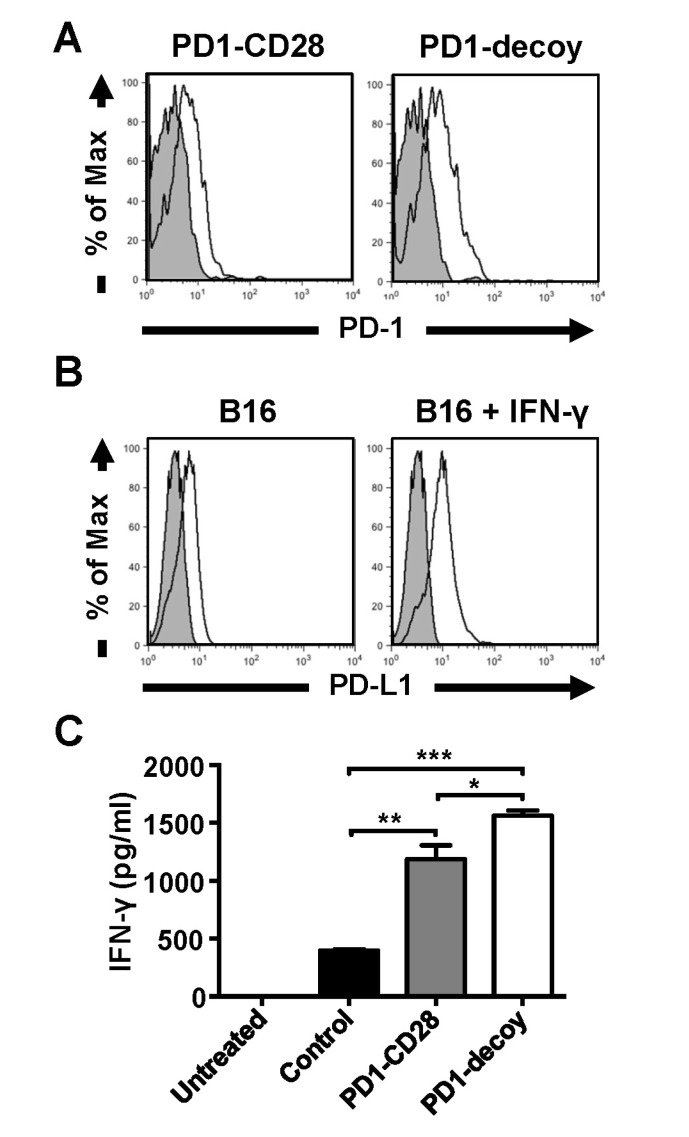
Figure 3. The PD-1-CD28 chimera does not improve the anti-tumor reactivity of T lymphocytes compared to PD-1 decoy. B16 melanoma antigen (gp100)-specific CD8 T cells (Pmel-1 cells) were transduced with retroviruses carrying PD-1-CD28 chimera or PD-1 decoy. (A) Expression levels of PD-1-CD28 chimera and PD-1 decoy in GFP-positive Pmel-1 populations were analyzed by flow cytometry. (B) B16 melanoma cells were either left untreated or treated with IFN-γ (20 ng/ml) for 48 hours. Then, PD-L1 expression was determined by flow cytometry. (C) Retrovirus-transduced GFP positive Pmel-1 cells were sorted by flow cytometry. The sorted cells (1×10⁵) were co-cultured with IFN-γ-treated B16 melanoma cells (1×10⁴) for 48 hours. IFN-γ in the cultured supernatants was quantified by ELISA (Student's t-test, *p<0.05, **p<0.01, ***p<0.001).

Figure 3