. 2016 Apr 28;16(2):99–108. doi: 10.4110/in.2016.16.2.99

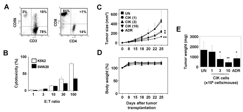
Figure 1. Antitumor efficacy of cytokine-induced killer (CIK) cells for CRC in a mouse xenograft model. CIK cells were generated by culturing human peripheral blood mononuclear cells (PBMCs) with a combination of IL-2 and anti-CD3 antibody for 14 days. CIK cells were stained with human monoclonal antibodies against CD3, CD56, CD4, and CD8 (A). In vitro cytotoxicity of CIK cells was evaluated by a 4-h 51Cr-release assay that used SW620 and K562 as target cells (B). Effector and target cells (1×10⁴ cells/100 µl/well) were mixed at different ratios (1:1 to 100:1). The percentage of cytotoxicity was calculated as following: cytotoxicity = [(sample-spontaneous)/(maximum-spontaneous)]×100. Spontaneous release was measured from target cells incubated in medium alone, whereas maximum release was measured after cells were treated with 2% Nonidet P-40. Nude mice (n=7) were implanted subcutaneously with 6×10⁶ SW620 cells. CIK cells (1 to 10×106 cells/mouse) were injected intravenously once a week. Adriamycin (ADR) was injected intravenously at 2 mg/kg. Tumor size was estimated as length (mm)×width (mm)×height (mm)/2 (C). Body weight was measured to estimate toxicity (D). Mice were sacrificed on day 25 and tumors were weighed (E). Statistical significance was determined by Student's t-test versus the PBS-treated control group (*p<0.05, **p<0.01). All experimental procedures were approved by the Animal Experimentation Ethics Committee and by the Institutional Ethics Committee of Chungbuk National University. Informed consents have been obtained from volunteers.

Figure 1