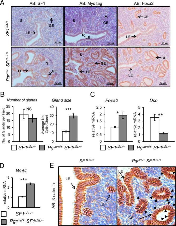
Figure 1.
Enhanced glandular development. A, Immunohistochemical staining for SF1, MYC-tag, and FOXA2 in representative uterine cross-sections of 8- to 10-week-old ovariectomized females. B, Quantification of glands and glandular size. C, RT-qPCR analysis for Foxa2 and Dcc message. D, RT-qPCR analysis for Wnt4 mRNA levels. E, Immunostaining of β-catenin. S, stroma; GE, glandular epithelium; LE, luminal epithelium. Two tailed t test significance indicated by ***, P < .001; **, P < .01; *, P < .05 and NS, not significant. SF1LSL/+ n = 8 and Pgrcre/+ SF1LSL/+ n = 6.

