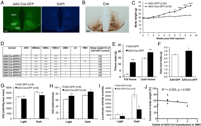
Figure 2.
Deletion of the Bdnf gene in the VMH of adult mice leads to obesity. A, Representative images showing AAV-Cre-GFP transduction in the Bdnflox/lox hypothalamus. DAPI staining was used to reveal anatomic structures. Scale bar, 250 μm. B, Cre immunoreactivity of brain sections from Bdnflox/lox mice injected with AAV-Cre-GFP into the VMH. Scale bar, 250 μm. C, Body weight of female Bdnflox/lox mice injected with either AAV-GFP (n = 8) or AAV-Cre-GFP (n = 8). Two-way ANOVA indicates a significant effect of AAV injection on body weight: F1,140 = 61.14, P < .0001. D, Analysis of injection sites in mice with AAV-Cre-GFP targeted to the VMH. The number of the * symbol indicates the extent of AAV transduction in different hypothalamic regions. E, Body composition of mice at 10 weeks after AAV injection (n = 8 mice for each treatment). Fat tissues were 8.6% and 29.8% of body weight in mice injected with AAV-GFP and mice injected with AAV-Cre-GFP, whereas lean tissues were 63.6% and 50.6% of body weight in mice injected with AAV-GFP and mice injected with AAV-Cre-GFP, respectively. F, Daily food intake of mice at 6 weeks after AAV injection (n = 8 mice for each treatment). G–I, VO2 and locomotor activity of mice at 10 weeks after AAV injection. J, Correlation between the extent of AAV-Cre-GFP transduction in the DMH and body weight at 9 weeks after injection. Error bars indicate SEM. LH, lateral hypothalamus; PMV, ventral premammillary nucleus; VMHc, central part of VMH; VMHdm, dorsomedial part of VMH; VMHvl, ventrolateral part of VMH.

