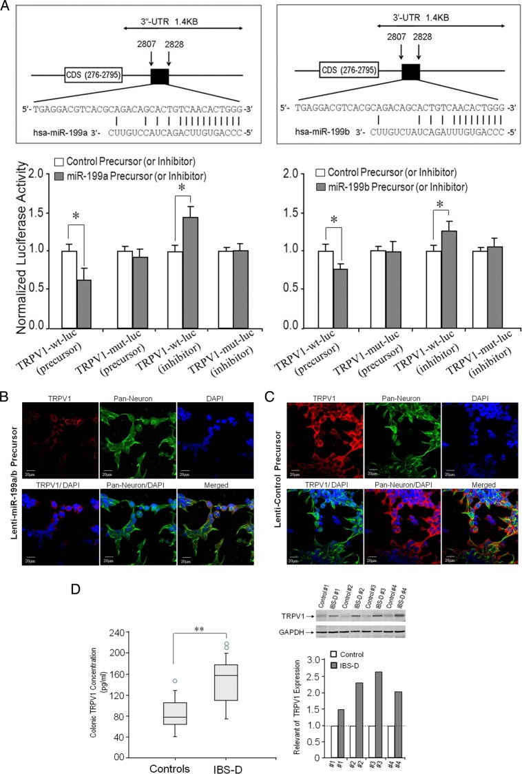
Figure 2.
Identification of transient receptor potential vanilloid type 1 (TRPV1) as a target gene for miR-199. (A) Upper panel: the location of the putative miR-199a/b target site in the TRPV1 3′-UTR is shown with highly conserved binding site for miR-199a/b in the 3′-UTR region of the TRPV1 mRNA, which codes for CDS 276–2795, a well-characterised capsaicin (vanilloid) receptor (upper panel left: miR-199a; upper panel right: miR-199b). (A) Lower panel: relative luciferase reporter co-transfected either TRPV1-wild type (wt) or TRPV1-mut (mutation) firefly luciferase expression construct, along with either miR-199a/b or control precursors (or inhibitors). Luciferase assays were done after 48 h using a dual luciferase reporter assay system. All the groups are normalised to the average of TRPV1 MUT group co-transfected either with control precursor or with control anti-microRNA (miRNA) (average value=1). A significant decrease (or increase) in relative firefly luciferase activity in the presence of wt miR-199a/b (lower left: miR-199a; lower right: miR-199b) with miR-199a/b precursors (or anti-miR-199a/b inhibitors) indicates the presence of a miR-199a/b modulated target sequence in the 3′-UTR of TRPV1. Bars show mean±SD from eight separate experiments done in triplicate (*p<0.05). (B and C) Confocal immunofluorescent staining of human neuron cells. TRPV1 was reduced after miR-199a/b overexpression (B) compared with control precursor (C). (D) Left panel: ELISA assay of colonic TRPV1 expression in patients with diarrhoea-predominant IBS (IBS-D). Higher levels of TRPV1 expression were found in colon biopsies from patients with IBS-D (n=45) compared with controls (n=40) (**p<0.01). The circles outside the boxes are outliers (data points >1.5*IQR). Right panel: Substantial increases in colonic TRPV1 expression in patients with IBS-D (n=4) compared with controls (n=4) on western blot analysis (right upper panel). We have quantitative bands from the western blots and analysed the data with an internal control of GAPDH (right lower panel). DAPI, 4′,6-diamidino-2-phenylindole; GAPDH, glyceraldehyde 3-phosphate dehydrogenase.

