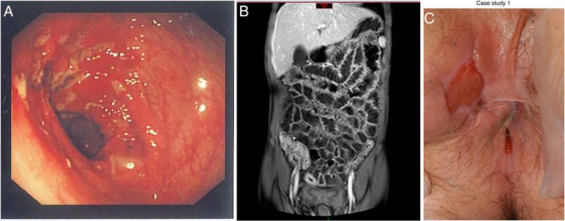
Figure 1.
(A) Left: endoscopic view of the transverse colon in Crohn’s disease (CD) revealing a cluster of deep ulcers. (B) Centre: magnetic resonance enterography showing extensive terminal ileal disease. (C) Right: perianal disease (inflamed skin tag and external orifice of a fistula at 10:00) in CD patient (reproduced with permission).

