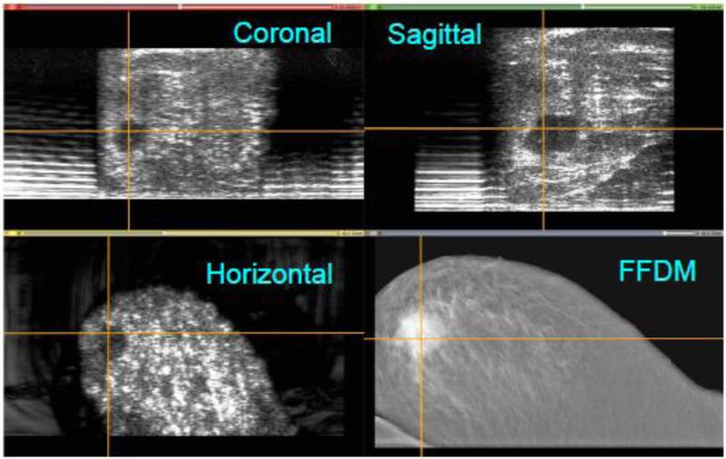
Figure 10.
Co-registration of the full-field digital mammogram (FFDM) in the horizontal plane and the automated breast ultrasound (ABUS) images in the horizontal, coronal and sagittal planes. A malignant lesion has been highlighted by cross hairs in the ABUS views, and is clearly is co-registered in the FFDM image. Note that for the ABUS images, the sagittal plane view is the acquired image, whereas the coronal and horizontal plane views have been reconstructed.

