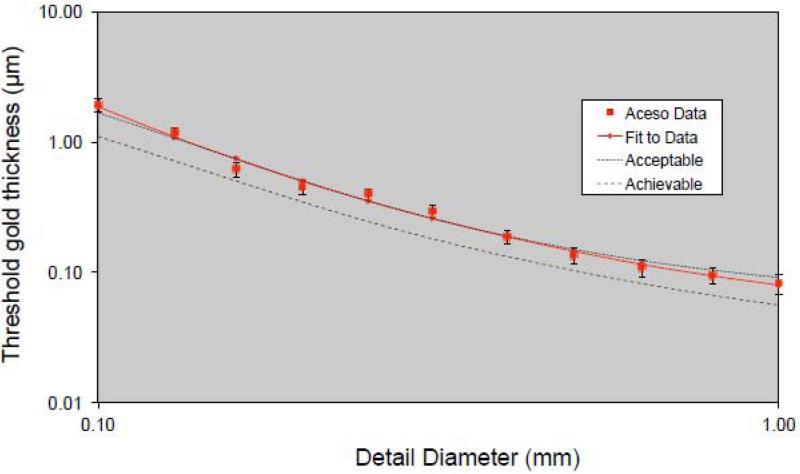
Figure 4.
Data for the CDMAM phantom generated by the EUREF software package (www.euref.org). The threshold gold thickness has been plotted as a function of detail diameter, with both axes using a logarithmic scale. The Aceso data are based on eight sequential X-ray images (at 31 kV, 27 mAs, 2.1 mGy) and may be compared with the EUREF standards of “acceptable” and “achievable” [37].

