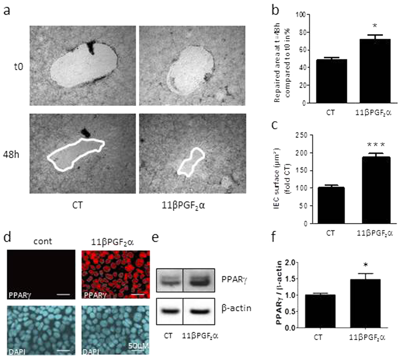
Figure 5. Functional impact of 11βPGF2α on IEB healing.
IEC properties were evaluated in Caco-2 monolayers after three days of culture in presence of lipid mediator 11βPGF2α or not (CT).(a) Representative pictures of epithelial healing of IEC monolayers cultured without treatment (CT) or with 11βPGF2α for two days. (b) Quantification of epithelial healing was calculated as a percentage of healing between t0 and 48 h. n = 5–12; Kruskal-Wallis test; *p < 0.05 as compared to IEC without treatment. (c) Epithelial spreading was expressed as percentage of CT IEC surface. n = 5–8 independent experiments performed in duplicates; Kruskal-Wallis test; ***p < 0.005 as compared to CT. (d) IEC properties were also evaluated in Caco-2 monolayers after two days of co-culture with Control EGC or CD EGC in presence of the lipid mediator 11βPGF2α or not (−). Quantification of epithelial healing was calculated as a percentage of healing between t0 and 48 h. n = 3; Kruskal-Wallis test; *p < 0.05 as compared to CD EGC-Caco2 co-cultures without treatment. §p < 0.05 as compared to Control EGC-Caco2 co-cultures without treatment. (e) PPARγ immunostaining on Caco-2 treated 5min with 11βPGF2α compared to ZO-1 staining. n = 3 independent experiments, scale 20 μM. (f) Western blotting analyzes of PPARγ expression in Caco-2 treated 24 hours with 11βPGF2α. (g) Quantification of PPARγ/β-actin related to the average control ratio taken as 1. Data represent mean ± SEM, n = 4 independent experiments; Kruskal-Wallis test; *p < 0.05 as compared to cont.

