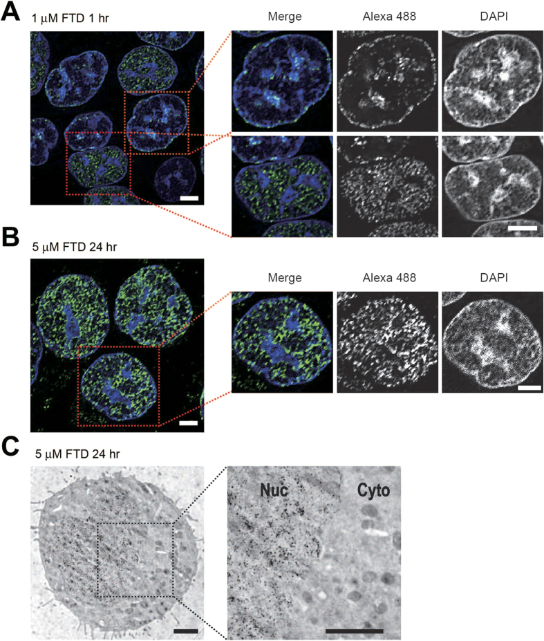
Figure 4. Immunodetection of FTD with the anti-BrdU antibody 3D4 at a high magnification.
(A,B) Immunofluorescence images of FTD acquired by confocal microscopy. HCT-116 cells cultured in the presence of 1 μM FTD for 1 hour (A) or 5 μM FTD for 24 hours (B) were immunostained with the anti-BrdU antibody 3D4 and an Alexa 488-conjugated secondary antibody. Nuclei were counterstained with DAPI. Typical 0.2 μm deconvolved images are shown. Scale bars indicate 5 μm. (C) FTD detection by immunoelectron microscopy. HCT-116 cells cultured in the presence of 5 μM FTD for 24 hours were immunostained using the anti-BrdU antibody 3D4 and a secondary antibody conjugated to gold particles with a diameter of 10 nm diameter. Scale bars indicate 2 μm.

