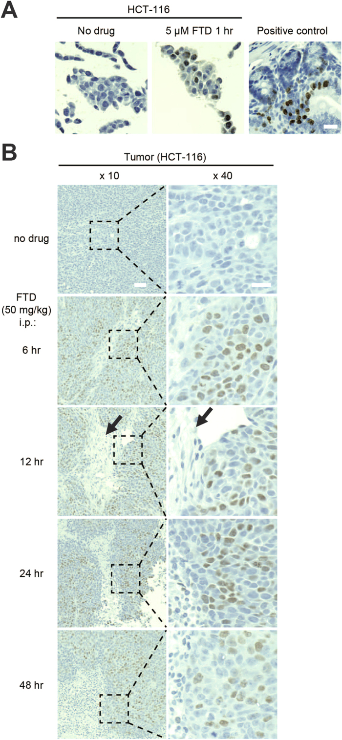
Figure 5. FTD detection of paraffin-embedded specimens by immunohistochemical staining.

(A) FTD detection in paraffin-embedded HCT-116 cells. HCT-116 cells were cultured in the presence or absence of 5 μM FTD for 1 hour. The cells were fixed with 10% formalin neutral buffer in PBS buffer, mixed with OCT compound, and embedded into paraffin. FTD was immunohistochemically stained with anti-BrdU antibody (3D4) using the BrdU in situ detection kit. Mouse intestinal crypts treated with BrdU were used as positive control. Scale bar indicates 20 μm. (B) FTD detection in paraffin-embedded HCT-116 xenografts in nude mice treated intraperitoneally with 50 mg/kg FTD. Scale bars indicate 50 μm (×10) or 20 μm (×40).
