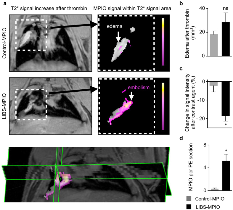
Figure 5. Contrast agent-induced signal voids can be detected within edematous area.
(a) In vivo
 weighted MRI of the thorax after intravenous application of human thrombin (left panel) and injection of Control-MPIO (right panel, above) or LIBS-MPIO (right panel below). Right panel show extracted overlay of edema (gray zone) and MPIO-induced signal decay within this area (pseudo-colored pink-yellow). Below, 3D map of edema after thrombin and corresponding signal overlay after injection of LIBS-MPIO in the murine lung. (b) Edema area after thrombin injection in LIBS-MPIO or Control-MPIO group does not differ (6 mice per group, ns = not significant). (c) Percent change of signal intensity in human thrombin-induced edema after injection of LIBS-MPIO or Control-MPIO (n = 6 mice per group, p < 0.05, Mann-Whitney U Test). (d) Mean number of MPIO found per immunohistochemistry section of pulmonary embolism after ex vivo preparation and staining for platelet CD41 (n = 6 per group, p < 0.05, Mann-Whitney U Test). Data are shown as mean ± s.e.m.

