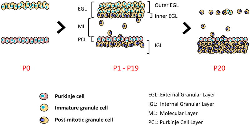
Figure 4. Postnatal cerebellum development involves the proliferation, migration and differentiation of granule cells.
(A) Following migration from the embryonic rhombic lip, granule precursor cells populate the external granular layer and engage in massive cellular expansion. (B) The initial proliferation stage of immature granule cells is closely followed by subsequent differentiation and inward migration of granule cells past the Purkinje cell layer (pink cells). Two sub-layers can then be distinguished: the proliferative outer layer (depicted with blue nuclei) and the differentiated inner layer (represented with purple nuclei). Post-mitotic granule cells migrate out of the external granular layer to invade and populate the internal granular layer (P1 to P19). (C) Around three weeks post-partum, all granule cells have left the external granular layer, completing the development of the cerebellum. The empty space between the external granular layer and the Purkinje cell layer is the molecular layer. It primarily contains the dendritic tree of Purkinje cells and axonal projections of different cell types of the cerebellar cortex: mature granule cells, Basket cells, Stellate cells, and Golgi cells. The cell bodies of Basket cells and Stellate cells are found in the molecular layer.

