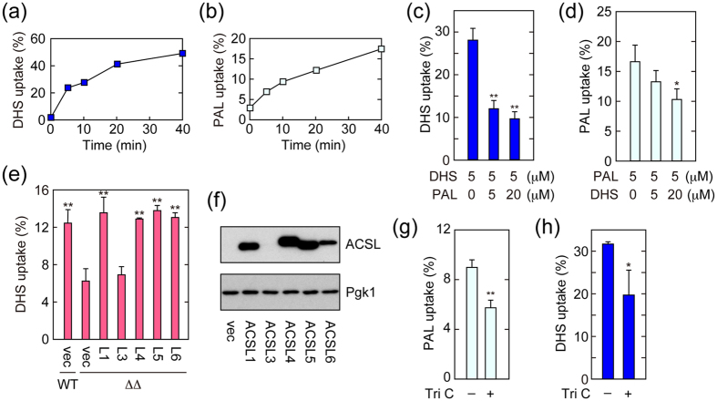
Figure 5. Mammalian ACSLs are involved in DHS uptake.
(a,b) IEC-6 cells were incubated with 5 μM [3H]DHS (a) or [3H]palmitic acid (PAL) (b) for the indicated time period. Radioactivities associated with cells and medium were counted by a liquid scintillation counter, and those associated with cells are expressed as a percentage of the total radioactivity. (c,d) IEC-6 cells were labeled with 5 μM [3H]DHS (c) or 5 μM [3H] palmitic acid (PAL; d) in the presence of cold palmitic acid or DHS, respectively, at the indicated concentration at 37 °C for 15 min. Transport activity was determined as in (a). Values represent the means ± SDs of three independent experiments, and statistically significant differences are indicated (t-test; *p < 0.05; **p < 0.01). (e,f) BY4741 (wild-type; WT) or AOY13 (faa1Δ faa4Δ; ΔΔ) cells harboring the pAKNF316 (vector; vec), pAO15 (3xFLAG-ACSL1; L1), pAO16 (3xFLAG-ACSL3; L3), pAO17 (3xFLAG-ACSL4; L4), pAO19 (3xFLAG-ACSL5; L5), or pAO21 (3xFLAG-ACSL6; L6) plasmid were grown in SC-URA medium at 30 °C. (e) Cells were labeled with 20 μM [3H]DHS at 30 °C for 5 min. Radioactivities associated with cells, medium, and glass test tubes were counted by a liquid scintillation counter, and those associated with cells are expressed as a percentage of the total radioactivity. Values represent the means ± SDs of three independent experiments. Statistically significant differences with respect to the value of AOY13 (faa1Δ faa4Δ) cells harboring vector are indicated (t-test; **p < 0.01). (f) Total cell lysates were prepared, separated by SDS-PAGE, and subjected to immunoblotting with anti-FLAG or, to demonstrate equal protein loading, anti-Pgk1 antibody. (g,h) IEC-6 cells were treated with DMSO or 20 μM triacsin C (Tri C) at 37 °C for 1 hr. Cells were then labeled with 5 μM [3H] palmitic acid (g) or 5 μM [3H]DHS (h) at 37 °C for 15 min. Transport activity was determined as in (a). Values represent the means ± SDs of three independent experiments, and statistically significant differences are indicated (t-test; *p < 0.05; **p < 0.01).

