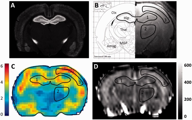
Figure 2.
Parallel anatomical, MRI, MRE and CBF maps of a juvenile rat brain section. (a) CB1R immunolabeling on a coronal section of a rat brain at postnatal day 11. (b) Right: High-resolution coronal T2-weighted MRI image. Left: Corresponding brain atlas map from Paxinos G and Watson C, exhibiting three hand-drawn regions of interest used to extract mean values from both MRE and CBF maps (1: hippocampus, 2: thalamus, 3: cortical grey matter). Ctx: Cortex; Thal: Thalamus, Hip: Hippocampus; MGP: Medial Globus Pallidus, Amyg: Amygdala. (c) MRE map of the absolute values (kPa) of the complex-valued shear modulus |G*|. (d) Cerebral blood flow map (ml/min/100 g).

