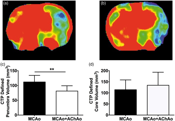
Figure 4.
Study II – CTP core and penumbra volume analysis in animals with or without concomitant AChAo during intraluminal MCAo. Representative 1-h CBF maps for animals with MCAo (a) or MCAo + AChA (b). Penumbra volume (c), core volume (d) at 1 h post-MCAo in animals with MCAo only (black bars) and animals with MCAo + AChAo (white bars). **p < 0.01. AChA: anterior choroidal artery; AChAo: anterior choroidal artery occlusion; CTP: computer tomography perfusion; MCAo: middle cerebral artery occlusion.

