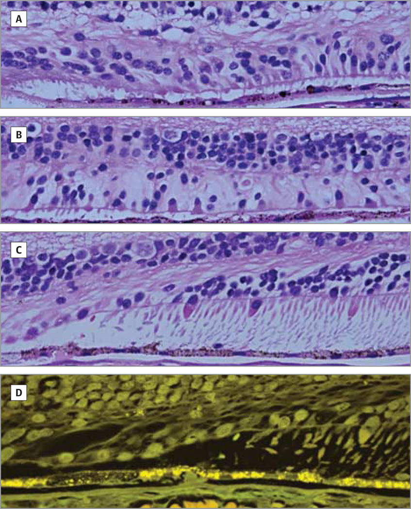
Figure 3. Light and Autofluorescence Micrographs.
There is variable loss of photoreceptor cells beyond the edge of geographic atrophy. Light (A–C) and autofluorescence (D) micrographs of an 84-year-old female donor in whom geographic atrophy had been diagnosed 3 years before death. A, At the edge of geographic atrophy, 1 to 2 layers of photoreceptor nuclei are seen. B, For up to 1200 µm from the edge, there are few cones with inner segments. Beyond 1200 µm, rods and cones with outer segments are seen (C and D), without there being a difference in the retinal pigment epithelium between that near the edge of geographic atrophy and that at 1200 µm away from the edge. The Bruch membrane is modestly thickened and the choriocapillaris is perfused. Original magnification ×2500.

