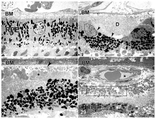
Figures 26–29.
Transmission electron micrographs of the retinal pigment epithelium (RPE)-choroid interface in eyes of donors without (Figure 26) and with (Figures 27–29) a clinically documented history of age-related macular degeneration (AMD). A single druse (D) is shown in Figure 27; its location between the RPE basal lamina (arrowheads) and Bruch’s membrane (BM) is clearly indicated. BLamD (asterisk) accumulates between the basal surface of the RPE and its basal lamina (arrow), whereas BLinD is located within the innermost aspect of Bruch’s membrane (Figure 28). A patent choroidal neovessel (asterisk), lying between BM and a layer of BLamD (rectangle), is shown in Figure 29. RPE=retinal pigment epithelium; BM=Bruch’s membrane.

