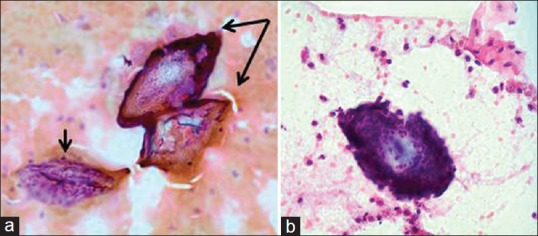
Figure 3.

(a) Cervical smear, magnification (Papanicolaou stain, ×40). Poorly preserved smears with cellular detail obscured by blood and inflammatory cells. Small arrow: Non-diagnostic, poorly preserved epithelial cells. Double arrow: Two Schistosoma haematobium ova, identifiable by their refractile shells and characteristic terminal spines. (b) Cervical smear, magnification (Papanicolaou stain, ×40). Schistosoma ovum surrounded by many neutrophil granulocytes. Terminal spine detected on fine focus, however, not seen clearly in this image
