
Figure 2.
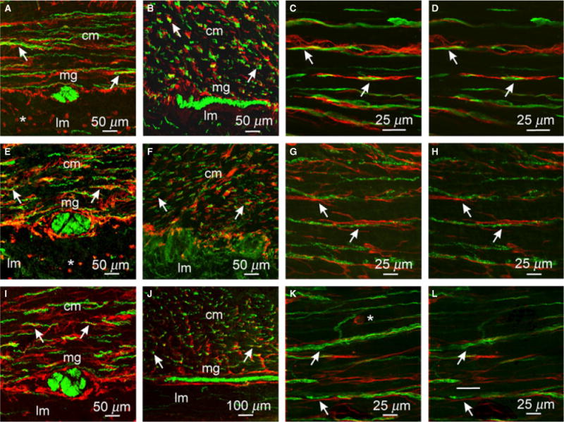
Close morphological relationship between enteric neurons and ICC in the gastric antrum. (A–D) Cryostat sections (A,B) and flat mount sections (C,D) of PGP9.5+ neurons (green) and Kit+ ICC (red). PGP9.5+ nerve fibers were observed in close association with ICC (arrows) in cryostat sections cut parallel (A) or transverse (B) to the CM and in flat mounts (C,D) within the CM (cm) but not the LM (lm). (E–H) Double labeling with sub-P and Kit in the antrum revealed a close anatomical relationship between excitatory nerve fibers and ICC. Cryostat sections cut parallel (E) and transverse (F) to the CM and flat mounts (G,H) cut parallel to CM fibers showed close apposition between sub-P containing neurons and ICC-IM (arrows). (I–L) nNOS neurons and ICC were also in close apposition to one another. (I, J) cryostat sections (K,L) flat mounts of nNOS (green) and Kit+ ICC (red) closely apposed to one another in the CM (cm; arrows). Both nerve fibers and ICC were sparse in the LM. (D,H,L) Single sections (0.4 μm) taken from the reconstructed stacks in C,G, and K, respectively. ICC-MY also surrounded myenteric ganglia (mg) but did not penetrate ganglionic sheaths. Numerous small rounded mast cells (*) were observed in the LM and occasionally within the CM. Scale bars are as indicated in each panel.