
Figure 5.
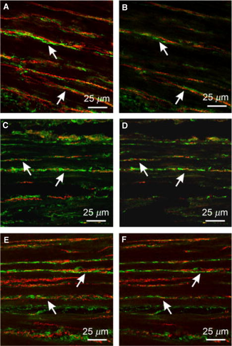
Relationship between enteric neurons and PDGFRα+ cells in the gastric fundus. (A,B) Double labeling with PGP9.5 and PDGFRα antibodies reveal close apposition between enteric neurons (red) and PDGFRα+ cells (green) in CM and LM. PGP9.5+ fibers ran parallel to PDGFRα+ cells for at least 130 μm. Double labeling with sub-P (C,D) or nNOS (E,F) reveal that both excitatory and inhibitory nerve fibers run parallel to PDGFRα+ cells for at least 150 μm. Scale bars are as indicated in each panel.