
Fig. 2.
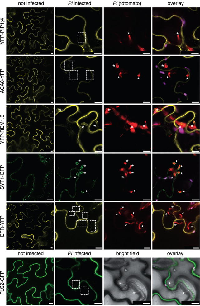
Plasma membrane-resident proteins differentially localize to Pi haustoria. N. benthamiana leaves transiently expressing the indicated fluorescently tagged proteins were infected with Pi 88069td or 88069 (lower panel) and imaged 3 dpi. Representative confocal micrographs show cross-sections of non-infected and Pi infected leaves. Pi haustoria are indicated by asterisks. The YFP–PIP1;4, ACA8–YFP EFR–YFP and FLS2–GFP signals are visible at the plant cell plasma membrane but not around haustoria (dashed boxes), while the YFP–StRem1.3 and SYT1–GFP signals are visible at haustoria. Bar = 10 μm.