
Fig. 4.
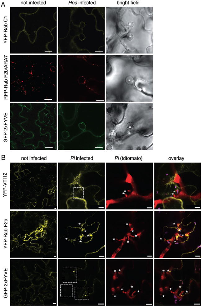
Endosomal compartments differentially localize around Hpa and Pi haustoria. A. Confocal micrographs of Arabidopsis transgenic lines expressing the indicated fluorophore fusions show cross-sections of non-infected and Hpa-infected leaves at 3 dpi. Hpa haustoria are shown in bright field images indicated by asterisks. YFP–Rab C1, RFP–ARA7 and GFP–2xFYVE signals are surrounding Hpa haustoria. Bar = 10 μm. B. Confocal micrographs of N. benthamiana leaves transiently expressing the indicated fluorophore fusions show cross-sections of non-infected and Pi-infected leaves at 3 dpi. Pi haustoria are indicated by asterisks. While YFP–Rab F2a accumulates around haustoria, YFP–RabC1 and GFP–2xFYVE signals are surrounding Pi haustoria but do not show a significant accumulation. Bar = 10 μm.