
Fig. 5.
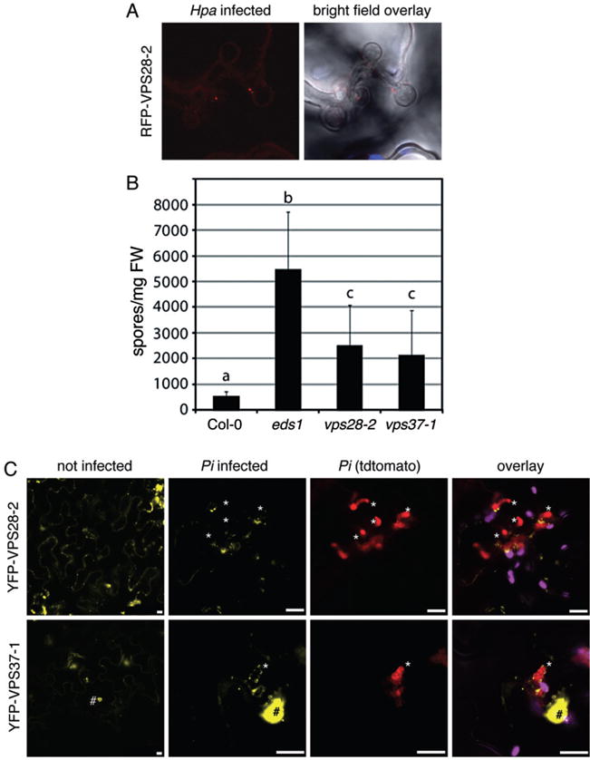
ESCRT-I components contribute to immunity against Hpa infection. A. Representative confocal images of RFP–VPS28-2 expressing plants show cross-sections of leaves infected with Hpa at 4 dpi. Hpa haustoria are shown in bright field images indicated by asterisks. RFP–VPS28-2 vesicles are present at Hpa haustoria. B. Spores of Hpa Waco 9 were quantified at 7 dpi on eight 2-week-old seedlings. Different letters indicate statistically significant of P <0.05 based on multiple pairwise comparisons based on standard post hoc ANOVA analysis. Error bars represent SD. C. Confocal micrographs of N. benthamiana leaves transiently expressing the indicated YFP fusions show cross-sections of non-infected and Pi-infected leaves at 3 dpi. Pi haustoria are indicated by asterisks. YFP–VPS28-2 and YFP–VPS37-1 positive compartments localize around Pi haustoria. YFP–VPS37-1 also localizes to subnuclear foci (#). Bar = 10 μm.