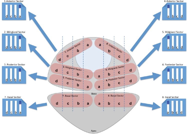
Figure 1. TPSB scheme used in our Institution. The biopsy starts from the right paraurethral region, sampling the anterior, mid, and posterior part of the gland. The ultrasound probe is then moved to show the most lateral part of the gland, which is again sampled in the anterior, mid and posterior regions. Next, the probe is moved medially and the regions of the gland comprised between the paraurethral and lateral region of the gland are sampled in their anterior, mid, and posterior part. The same procedure is performed on the left side.

