Figure 3.
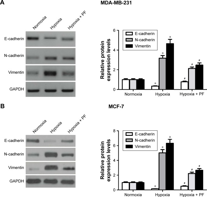
PF prevented the hypoxia-induced EMT in breast cancer cells.
Notes: (A) MDA-MB-231 cells grown under normoxia and hypoxia were treated with 25 μM PF for 24 hours. E-cadherin, N-cadherin, and vimentin expressions were assessed by Western blot analysis. The relative protein expression levels of E-cadherin, N-cadherin, and vimentin were quantified using Image-Pro Plus 6.0 software (Media Cybernetics, Silver Spring, MD, USA) and normalized to GAPDH. (B) MCF-7 cells grown under normoxia and hypoxia were treated with 25 μM PF for 24 hours. E-cadherin, N-cadherin, and vimentin expressions were assessed by Western blot analysis. The relative protein expression levels of E-cadherin, N-cadherin, and vimentin were quantified using Image-Pro Plus 6.0 software and normalized to GAPDH. Each bar represents the mean ± SD. The results were reproduced in three independent experiments. *P<0.05 vs normoxia group, #P<0.05 vs hypoxia group.
Abbreviations: PF, paeoniflorin; EMT, epithelial–mesenchymal transition; SD, standard deviation.

