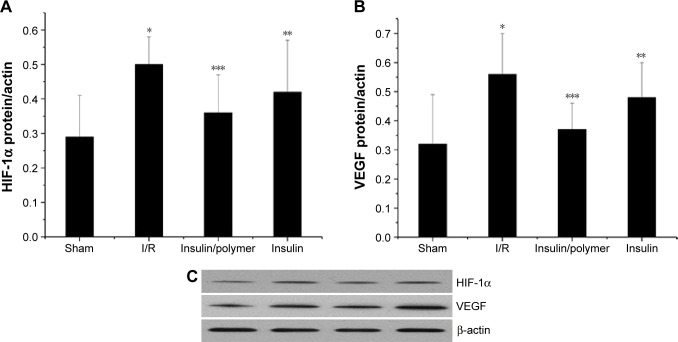
Figure 10.
The qualitative expression (C) and quantitative analyses of HIF-1α (A) and VEGF (B) in pulmonary tissues for different groups of rats.
Notes: The pulmonary tissues of Sham, I/R, Insulin, and Insulin/polymer groups rats were collected 24 hours after reperfusion and the expression of HIF-1α and VEGF measured. Results are expressed as mean ± SD. (A) A significant increase from sham group is denoted by *P<0.01, a significant decrease from I/R groups is denoted by **P<0.01, and a significant decrease from I/R groups is denoted by ***P<0.01. (B) A significant increase from sham group is denoted by *P<0.01, a significant decrease from I/R groups is denoted by **P<0.01, and a significant decrease from I/R groups is denoted by ***P<0.01.
Abbreviations: HIF, hypoxia-inducible factor; VEGF, vascular endothelial growth factor; I/R, ischemia/reperfusion; SD, standard deviation.

