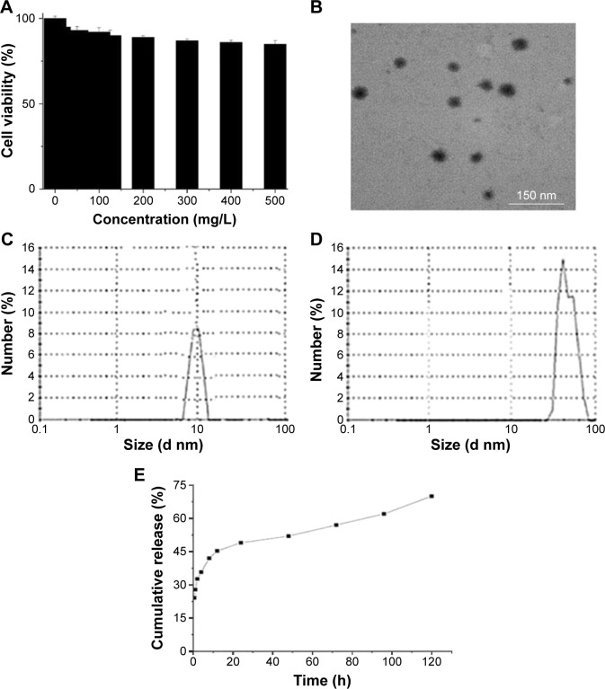
Figure 5.
Characterization of PEG-b-(PELG-g-PLL) and insulin/PEG-b-(PELG-g-PLL) complexes.
Notes: (A) Cellular viability of EC9706 cells cultured with different concentrations of PEG-b-(PELG-g-PLL); (B) TEM image of insulin/PEG-b-(PELG-g-PLL) complexes; (C) Diameter of block copolymer PEG-b-(PELG-g-PLL) in PB; (D) Diameter of insulin/PEG-b-(PELG-g-PLL) complexes in PB; (E) Cumulative releasing profile of insulin from insulin/PEG-b-(PELG-g-PLL) complexes.
Abbreviations: TEM, transmission electron microscopy; PB, phosphate buffer; PEG-b-(PELG-g-PLL), poly(ethylene glycol)-b-(poly(ethylenediamine l-glutamate)-g-poly(l-lysine)).

