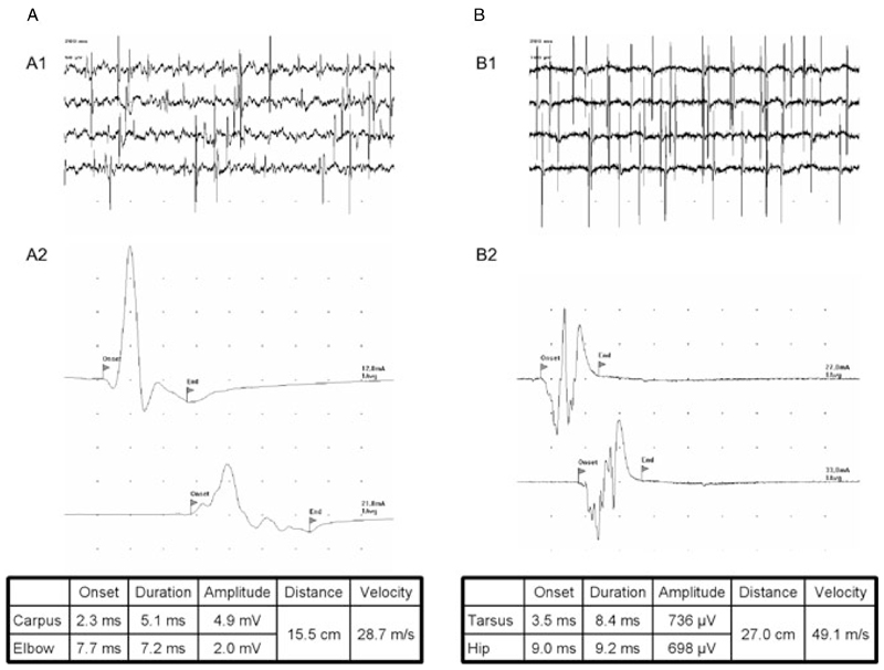
Figure 1.
Examples for results obtained in electrophysiologic investigations of acute canine polyradiculoneuritis (ACP) dogs (both dogs exhibited anti-GM2 Abs). (A) Dog 15; (B) dog 16. A1/B1, spontaneous activity in electromyographic assessments of the tibial cranial muscle; A2/B2, motor nerve conduction studies of ulnar (A2) and sciatic/tibial (B2) nerve. Note the reduction of CMAP-amplitude and CMAP dispersion in the second trace of dog A. This dog also exhibited a reduced motor nerve conduction velocity (MNCV; 28.7 m/s). Dog B exhibited a vast reduction of its CMAP amplitude in both traces (<1 mV) with a MNCV (49.1 m/s) at the lower end of the physiological reference rage. Divisions on the abscissa are 2 ms (A2) and 5 ms (B2). Divisions on the ordinate are 1 mV (A2) and 200 µV (B2).

