
Figure 3.
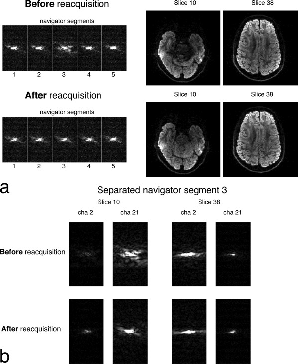
Navigator‐based reacquisition with two simultaneously excited slices (labelled with their anatomical slice number) with b = 1000 s/mm2 diffusion‐weighting. a: Before reacquisition, corruption in segment 3 is indicated by the dispersed k‐space in the navigator, which contains combined data from the two slices. When segment 3 is reacquired, artifacts are removed from the images. b: Single‐channel maps (all at the same scale) of the unaliased k‐space from navigator segment 3. Two channels are chosen which are close to slice 10 (channel 21) and 38 (channel 2) to demonstrate the variation in signal.