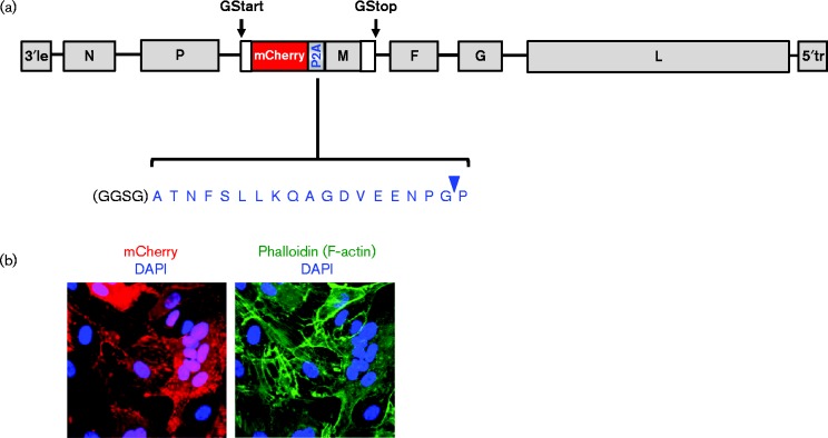
Fig. 1.
Schematic of recombinant NiV genome with the mCherry reporter expressed in the same transcriptional unit as the matrix (M) gene (rNiV-mChM). (a) mCherry expression is driven by the gene start (GStart) and gene stop (GStop) signals that flank the M ORF. mCherry and M are expressed as individual proteins via the P2A ribosomal skipping sequence (blue letters). Arrowhead (blue) indicates the ‘cleavage’ site between the glycine (G) and proline (P) residues. The M protein thus retains an additional N-terminal proline residue. For rNiV-mChM-opt, a GSG linker was inserted preceding the P2A sequence, with an additional glycine to preserve the rule of six for paramyxovirus genomes. (b) Human umbilical vein endothelial cells (HUVECs) were infected with rNiV-mChM and imaged by confocal microscopy. Left panel, mCherry can be seen in multinucleated syncytia caused by NiV-induced cell–cell fusion. Right panel, same field stained with phalloidin to detect F-actin for cell demarcation.

